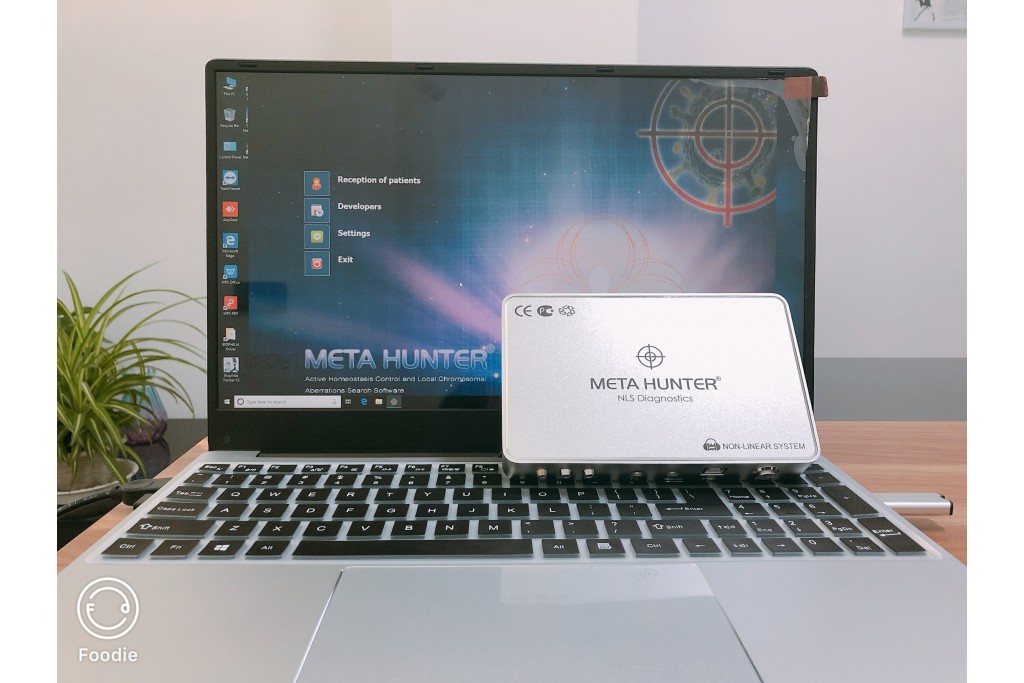
NLS Scanner with Metatron Hunter 4025 and Diagnosis of Muscle Trauma
- Wendy
- June 06, 2023
- 2206
- 0
- 0
According to the injury mechanism, lower extremity muscle injuries can be divided into three categories.
1. Damage caused by overwork. Strong start-up stresses, especially when insufficiently warmed up and too cold, can cause cracking. Also with overexertion, muscle overstretching can occur which in turn can cause pain or damage to the fascial compartment, which is most typical for the adductors of the hip and the posterior muscles of the buttock.
2. Actual trauma from external disruptive factors - direct or indirect (fall) blows.
3. Trauma developed as a result of sustained overload, «chronic microtrauma».
According to J. Comtet and W. Muller, complete ruptures occur in muscles with isolated functions. In the quadriceps of the thigh is the m. Rectus femoris without any synergists. This trauma occurs more often in hockey players after a hockey stick strike. The most frequent site of injury to the rectus femoris is at the tendon transition. Partial ruptures are more common in the biceps thigh and adductor thigh muscles. The integrity of the adductor muscle is compromised not only at the muscle-to-tendon transition but also at its attachment to the pubic bone. In the medical literature, this trauma is called «ARS-complex» (Adductor Rectus Syndrome). As the term implies, with an injury to the adductors, the rectus abdominus is injured at its junction with the syndesmosis.
Conditional muscle damage can be divided into microbursts (affected area 3-5mm) and ruptures greater than 5mm. Trauma can be longitudinal - along the muscle fibers, or transverse. Longitudinal trauma is more common and easier to diagnose and treat. Hematomas and synovomas visualized at trauma sites by NLS graphics. Diagnosis is complicated by NLS images showing polymorphism due to atypical traction and muscle ischemia at lateral bruises and ruptures.
Application of NLS scanner with Metatron Hunter 4025 allows to visualize muscle damage in sports trauma with high precision. The following signs are typical.
1. NLS-ultramicroscopic scan showing abnormal structural properties (streaks) of the wounded area, with moderately hyperchromic linear structures (5-6 points on the Fleindler scale) corresponding to myofibril bundle sheaths seen.
2. Hyperchromatic and metachromatic areas of various sizes and indistinct outlines present - hematomas. NLS-angiography plays an important role in its diagnosis. NLS-ultramicroangiography reveals the influence of vessel walls in these structures. This sign allows to distinguish between hematoma and histological process.
3. Pathological traction in the lesion area is more pigmented (5-6 points on the Fleindler scale) than intact muscle tissue.
During the comprehensive diagnosis, it is necessary to check the state of muscle tension and relaxation. It is recommended to measure the exposed pathological area for subsequent dynamic control.
Consequently, most traumas (94.5%) presented as muscle bruises smaller than 20 mm, difficult to diagnose by other means. At the same time, the wrong medical strategy and the wrong choice of training mode under such trauma can lead to further muscle damage, thereby limiting physical activity. This fact illustrates the importance of NLS research in the diagnosis of muscle trauma in athletes. At present, more and more athletes or families are beginning to use it, and Metatron Hunter 4025 appears more and more frequently in people's lives.

